Bioelectric Control of Axial Regeneration in Planaria


Model assumptions and mechanisms

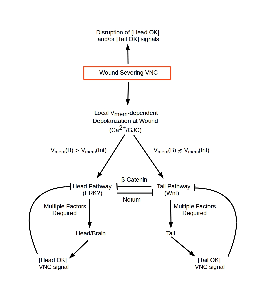
This model describes regeneration following amputations transverse to the anterior-posterior (A-P) axis in Planaria, i.e. amputations that sever both branches of the ventral nerve cord (VNC). Both single transverse wounds that remove anterior (head) or posterior (tail) structures and pairs of wounds that remove both anterior and posterior structures are considered. The model is based on four assumptions, two concerning the encoding of A-P polarity and two concerning the behavior of cells at the wound blastema:
  1. That A-P polarity information in planaria is encoded bioelectrically as an A-P polarization profile;

  2. That the A-P polarization profile in wild-type (WT) planaria is maintained by the tonic exchange of signals, here called [Head OK] and [Tail OK], between anterior (Brain) and posterior (VNC Terminus) ends of the VNC.

  3. Anterior and posterior wound blastema have identical differentiation potential and respond to all signals in the same way; and

  4. All cell-cell communication, including all bioelectric communication, is local.
The model considers two wound-response mechanisms as shown in the diagram on the right. Severing the VNC causes two immediate responses: Cells near the wound are assumed to respond to increased [Ca2+] by opening Ca2+ channels. This initial wound response is assumed to depend on the target-cell Vmem: relatively depolarized cells respond more strongly and open more Ca2+ channels, while relatively hyperpolarized cells respond less strongly and open fewer Ca2+ channels. Hence the net effect of increased [Ca2+] at the wound is to amplify differences in Vmem along the A-P axis of the amputation fragment. This amplification response is assumed to be dependent on gap junction communication (GJC) between cells. While the nature of the Ca2+ channel-opening signal is not yet characterized, the sensitivity of transverse wounds in planaria to local [Ca2+] is well documented (Beane et al. (2011) Chem. Biol. 18, 77-89; Chan et al. (2014) PLoS Path. 10, e1003942; Chan et al. (2017) Biochim. Biophys. Acta 1864, 1036-1045).

Cells at the wound blastema are assumed to measure the difference ΔVmem between their own membrane potential Vmem(B) and the average of the membrane potentials of their neighbors interior to the wound, Vmem(Int), which serves as a voltage reference. Following an anterior transverse wound, the neighboring cells that implement the local Vmem(Int) reference are all posterior to the wound blastema; following a posterior wound, the neighboring cells that implement the local Vmem(Int) reference are all anterior to the wound blastema. Each blastema has, therefore, an intrinsic bioelectric polarity.

Blastema cells are assumed to execute the differentiation pathway shown in the diagram to the right to produce either a head or a tail with its associated neural structures, i.e. a brain or a VNC terminus that re-integrate with the remnant VNC. The mutual inhibition implemented by β-Catenin and Notum is assumed to act downstream of the Head-Tail differentiation decision. The role of this mutual inhibition is to enforce a winner-take-all decision at each wound surface to implement either the head or the tail pathways, i.e. to prevent mixed outcomes in which some blastema cells choose the head pathway and some choose the tail pathway. This mutual inhibition is assumed to be Vmem sensitive, with expression of Notum turned off when depolarization is large and the tail pathway and hence β-Catenin expression is fully inhibited.

This version of the simulator does not include details of the molecular pathways implementing either the head or tail regeneration pathways. Completion of head and/or tail regeneration and re-integration of the VNC is assumed to induce the tonic "Head OK" signal from the brain and "Tail OK" return signal from the VNC terminus, completing the pathway

Using the model to simulate A-P axial amputation experiments in planaria

The model simulation tool below focuses on the initial choice point between head and tail pathways and calculates the branching ratio. A significantly positive ΔVmem (i.e. depolarization of the wound surface relative to the local interior reference) is assumed to initiate the differentiation of head structures and a near-zero or negative ΔVmem (i.e. neutrality or hyperpolarization of the wound surface relative to the local interior reference) is assumed to initiate the differentiation of tail structures. The model has three kinds of parameters: cut-position parameters, ΔVmem detection sensitivity parameters, and polarization parameters.

Intact wild-type (WT) worms are assumed, consistent with experimental measurements, to have an asymmetrical U-shaped Vmem profile along the A-P axis, with the head significantly more depolarized than the tail. This WT profile is represented by a "bathtub" sum of decreasing and (offset) increasing exponentials with equal decay rates but distinct coefficients. The cut-position parameters determine the wound and reference potentials for a post-amputation fragment given this initial Vmem profile. An anterior cut at 20% of intact-animal length is assumed to remove just the head, a 40% cut is immediately before the pharynx, a 60% cut is immediately after the pharynx, an 80% cut is midway between the pharynx and the tail tip; similarly for posterior cuts.

The ΔVmem detection sensitivity parameters determine how the polarization difference between the cells at the cut location and their local reference cells (i.e. the local ΔVmem) affects the probability of branching to the tail pathway. The "expectation" parameter sets the minimum polarization difference below which blastema cells execute the tail pathway: when the local ΔVmem is less than (equal to) this expectation value, the tail pathway branching ratio is greater than (equal to) 50%. The "precision" parameter sets the midpoint slope of the sigmoidal branching-decision function. It represents the resolution or accuracy with which blastema cells can measure ΔVmem.

The polarization parameters allow modifications of the polarization profile of the amputation fragment. Opening of Ca2+ channels in response to wounding is modelled by an an amplification of the fragment Vmem profile to reproduce the profile of the intact animal; this models the rapid recovery of depolarization, particularly at anterior wounds, observed experimentally. With all other parameters at default values, an amplification of 47% is required for 100% WT regeneration of middle fragments; for "pre-tail" (PT) fragments cut at 60% and 80% of adult length, the amplification needed for 100% WT regeneration is 82%. Wounds can also be depolarized or hyperpolarized to model the results of drug treatments, e.g. nigericin, that affect polarization at wound surfaces. With other parameters at default values, 40% depolarization of middle fragments produces 100% two-headed regenerates, while 10% hyperpolarization produces 100% two-tailed animals.

Step 1: Set initial Vmem profile in intact animal

The curve shown represents the relative density of depolarized cells and hence the neighborhood-average Vmem profile in mV along the A-P axis. It assumes a "bathtub" sum of exponentials with equal decay rates. The only parameter is the ratio of the coefficients of the two exponentials.

Set initial A-P average Vmem profile


Vmem(mV)                                   

Your browser does not support the HTML5 canvas tag.
Length of the intact worm


Step 2: Set model parameters and select amputation experiment

To initiate a model run, set the parameter values (or use the default values given) and select an amputation experiment. The model calculates and displays wound-surface potentials, local anterior and posterior reference potentials, and a midpoint reference potential for the selected amputation fragment (anterior, middle or posterior) using the Vmem profile specified above. The then calculates and displays pathway activation levels, and head and tail outcome probabilities. It also calculates the probabilties for "blob" outcomes that represent failure of either head or tail differentiation pathways.


Set parameter values

Set anterior cut positionSet posterior cut position
  
Set ΔVmem ExpectationSet ΔVmem Precision


Select amputation experiment


Your browser does not support the HTML5 canvas tag.

Initial amputation-fragment polarization profile:

Anterior, Anterior Reference      Midline Reference      Posterior Reference, Posterior

Your browser does not support the HTML5 canvas tag.


Step 3: Modify fragment polarization profile

The sliders below allow the initial fragment polarization profile to be modified in one three ways. Measurements of Vmem at wound sites of "middle" fragments indicate a rapid re-establishment of dense depolarization at anterior wounds and suggest a smaller re-depolarization at posterior wounds. This is consistent with Vmem dependent opening of Ca2+ channels in cells at the wound(s). "Amplify fragment polarization" models this process by effectively "stretching" the fragment by the selected amount to re-establish the whole-animal polarization density profile.

"Depolarize wound(s)" and "Hyperpolarize wound(s)" model drug experiments in which one or both wounds are artificially depolarized are hyperpolarized. The drug treatment is assumed not to extend significantly beyond the wound surface; hence these manipulations do not modify the local reference polarizations.

Amplify fragment polarizationDepolarize wound(s)Hyperpolarize wound(s)


Modified polarization model:

Anterior, Anterior Reference      Midline Reference      Posterior Reference, Posterior

Your browser does not support the HTML5 canvas tag.


 Anterior  Posterior 
HeadsBlobsTailsHeadsBlobsTails


Copyright © 2018 Chris Fields
Non-commercial re-use permitted; please cite:
Durant et al., in review.